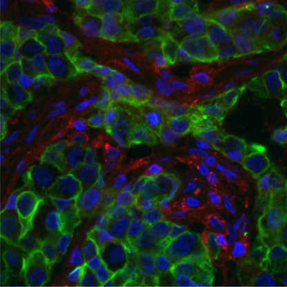
IF on FFPE Sections
(A) Deparaffinize slides: Xylene (2x5 min), 100% EtOH (2x3 min), 95% EtOH (1 min), 80% EtOH (1 min), ddH2O (≥ 5 min)
(B) Staining
- Antigen retrieval (select one)
- EDTA buffer
- 1 mM EDTA + 0.05% Tween 20 in DI water, pH 8.0
- Immerse slides in EDTA buffer in slide container
- Place slide container in a steamer for 30 minutes
- Cool for 30 minutes in the same buffer
- Citrate buffer
- 10 mM sodium citrate + 0.05% Tween 20 in DI water, pH 6.0
- Immerse slides in citrate buffer in slide container
- Place slide container in steamer for 30 minutes
- Cool for 30 minutes in the same buffer
- Proteinase K
- Apply Proteinase K solution for 30 s
- Rinse slides
- Wash 2x5 minutes in PBS
- Using PAP pen, draw around sections
- Block the section for 1 hour in blocking buffer (5% fetal horse serum, 1 mg/mL BSA, 0.05% Tween 20, 1X PBS) in a humidified chamber at RT
- Remove the blocking buffer
- Add the primary antibody, diluted to the desired concentration in blocking buffer. Incubate 1.5 hours at RT in humidified chamber (or overnight at 4°C)
- Remove the primary antibody
- Wash slides 3x 5 minutes in 1x PBS
- Re-block for 10 min
- Dilute secondary antibodies in blocking buffer and incubate for 1 hr at RT.
- Wash 1x 5min in PBS
- Add Hoeschst 33342 at 1/10,000 for 15 min.
- Wash 3x 5 min in PBS
- Carefully mount tissue on glass slides with ~2 drops of Prolong Gold mounting medium. Incubate overnight at RT in the dark to cure.
- Seal the edges with nitrocellulose based lacquer (nail polish)
- Mounted tissue should be stored protected from light at 4ºC and should be imaged as soon as possible to obtain best imaging results
- Analyze
Note: FFPE tissues have higher background in the green channel. A highly expressed antigen or use of a Brilliant Blue 515 secondary is recommended to compensate for this.
